Transient Muscle Paralysis Model
Using Botox as a paralyzing agent, we have developed a new model of disuse. in humans, disuse (or lack of skeletal loading) causes profound degradation of muscle and bone mass (e.g., exposure to microgravity, bed rest, or spinal cord injury). Botox (Botulinum neurotoxin A) is a neuromuscular blocking agent produced by Clostridium botulinum bacterium that can induce temporary muscle paralysis after intramuscular injection by blocking the release of acetylcholine into the neuromuscular junction (1).
In our initial studies, we used high resolution microCT imaging to assess bone morphology changes following transient muscle paralysis induced in the calf and quadriceps muscle groups by a single injection of Botox (Figure 1). Compared to saline injected controls, by 3 weeks we observed a profound degradation of trabecular bone in the proximal tibia metaphysis (-54.3%) and cortical bone at the tibia mid-diaphysis (-14.6%; Figure 2). Follow-on studies have indicated that the acute bone loss observed in this model is driven almost entirely by osteoclastic bone resorption (both for trabecular and cortical bone). Our current studies with this model are focused on understanding the signaling pathways responsible for inducing such rapid osteoclastogenesis.
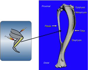
Figure 1: Botox or saline is injected into the calf and quadriceps muscle groups. We evaluate cortical bone in the tibia diaphysis and trabecular bone in the proximal tibia metaphysis using micro computed tomography imaging.
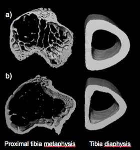
Fig. 2. MicroCT images (3D) of the right proximal tibia metaphysis and right tibia mid-diaphysis from a representative saline injected (a) and Botox injected mouse (b). Across groups, trabecular BV/TV (%) was reduced −54.3% at the proximal tibia, and cortical bone was reduced −14.6% at the tibia mid-diaphysis (all P < 0.001). Image from: Warner SE et al. Bone 2006;38:257-264.
Tracking Osteoclast Activity Using Serial microCT Image Registration
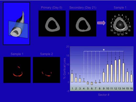
The goal of this study is to track the onset and migration of bone loss induced by transient muscle paralysis with the hopes of using this data to elucidate the drivers of osteoclastic bone resorption in this model. Bone loss induced by transient muscle paralysis is an osteoclast driven process that results in dramatic endocortical resorption at the tibia midshaft, while leaving the periosteal surface relatively unchanged. This unchanged periosteal surface can be used as a registration landmark to superimposed serial micro-CT images as a way to track the spatial and time dependent resorption that occurs on the endocortical surface (seen below). 3-Dimensional videos of bone loss seen at the bottom of this page, highlight the complex, yet highly reproducible spatial pattern of cortical bone loss observed in our model.
1. Hambleton P. Clostridium botulinum toxins: a general review of involvement in disease, structure, mode of action and preparation for clinical use. J Neurol. 1992 Jan;239(1):16-20.