Home
-
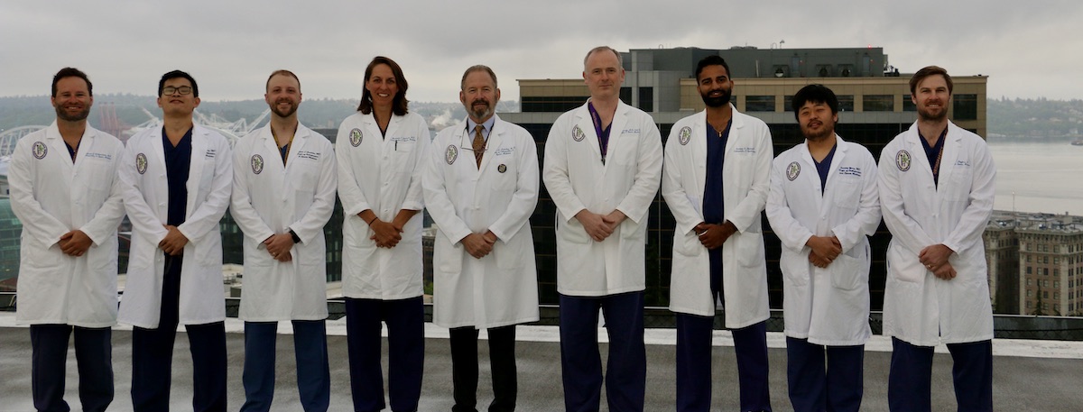
Our 2023 Graduating Residents
Congratulations and thank you for your hard work!
Learn More about our Graduates -

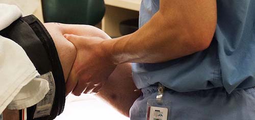
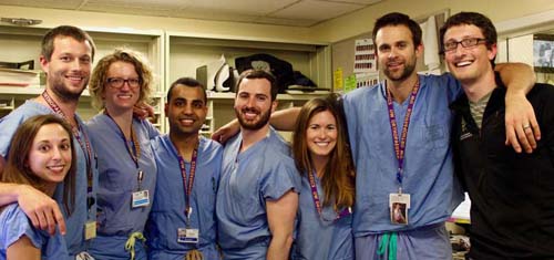
Our Residency Program
Dedicated to teaching in the context of excellence in patient care, we are preparing the next generation of leaders in orthopaedic surgery.
Learn More -
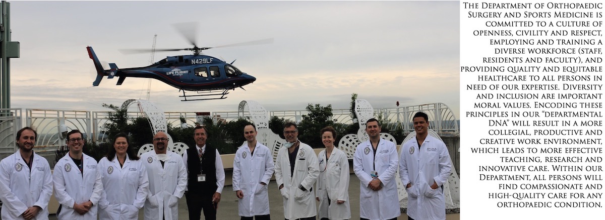
We are committed to a culture of openness, civility and respect, employing and training a diverse workforce (staff, residents and faculty), and providing equitable healthcare to any person in need of our expertise.
Learn More

The University of Washington Department of Orthopaedic Surgery and Sports Medicine is committed to a culture of openness, civility and respect, employing and training a diverse workforce (staff, residents and faculty), and providing quality and equitable healthcare to all persons in need of our expertise.

The University of Washington, Department of Orthopaedic Surgery and Sports Medicine is committed to improving diversity not only in our department but in our orthopedics community as a whole. Learn more at the links below about our efforts to improve diversity and inclusivity in our department and become leaders at UW Medicine and in the orthopaedics community to better reach under-represented students.
News
Videos
See More Videos